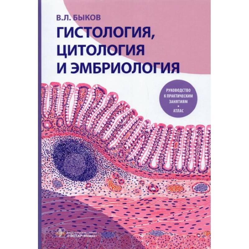
Histology, cytology, and embryology. Practical guide. Atlas: Textbook
Please sign in so that we can notify you about a reply
The purpose of this textbook is to promote the successful development of the material of practical classes in the study of the course of histology, cytology and embryology at a medical university. The book presents recommendations on the procedure for working with specific drugs and electronic micrographs for each study topic and their detailed descriptions are given with clarification of the characteristics of the structural organization and the function of individual components of cells, tissues and organs. At the same time, attention is paid to the most important cytological and histological details, the recognition of which serves as the basis for the identification of the drug or electron micrograph. Recommendations for the detection of these structural components in the studied drugs are given and their characteristics are considered. The description is illustrated by original drawings from histological preparations and electronic micrographs that reflect the main sections of the standard course on the subject. Each drawing is equipped with a detailed signature with designations.
The manual will help students master the necessary practical skills and competencies when working with histological drugs, electronic microphics and their digital images, as well as develop the skills of self -recognition of organs, tissues and cells, identifying various structural components and the functional interpretation of their condition.
The publication is intended for students of medical universities, clinical residents, graduate students, histologists, embryologists, doctors of various specialties.
It is recommended for students studying in the specialties on 05/31/01 Medical Affairs, 05/31/02 Pediatrics, 32.05.01 “Medical and preventive business”, 05/31/03 “Dentistry”
Author:
Author:Bykov V.
Cover:
Cover:Hard
Category:
- Category:Medical Books
Publication language:
Publication Language:Russian
Paper:
Paper:Molded
ISBN:
ISBN:978-5-9704-5225-7
No reviews found
