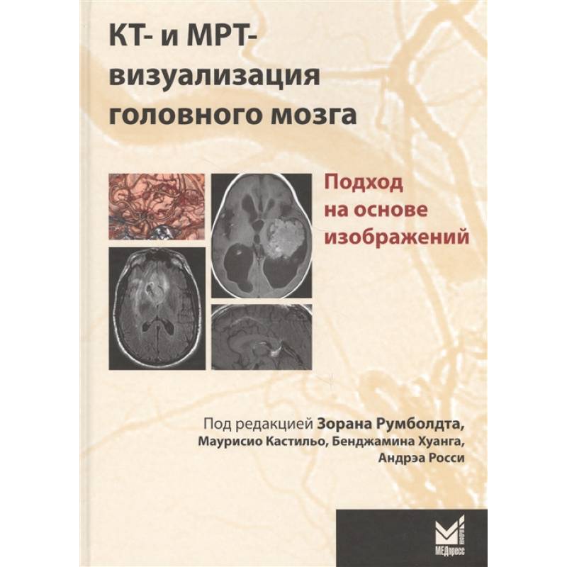
CT (computed tomography) and MRI (magnetic resonance imaging) are used for visualizing the brain. Image-based approach
Please sign in so that we can notify you about a reply
The book is built on the principle of the directory of differential diagnostics, the basis of its heading is the features of diagnostic images. Illustrations are placed on the strokes on the left, on the right is a capacious description of the observed pathology and a list for differential diagnosis indicating the pages on which differentiated conditions are described. The management gives more than 1,500 images (mainly computer and magnetic resonance tomograms) of the brain observed in more than 200 diseases. The book begins with a description of the picture of bilateral symmetrical lesions and median defects, since they are easiest to confuse with each other, especially if the reader has a relatively small experience.
The publication is intended for neuroralologists practicing radiologists of a common profile, neurologists, neurosurgeons, pediatricians, students of medical universities and faculties
The publication is intended for neuroralologists practicing radiologists of a common profile, neurologists, neurosurgeons, pediatricians, students of medical universities and faculties
Author:
Author:Румболдт З.
Cover:
Cover:Hard
Category:
- Category:Medical Books
Publication language:
Publication Language:Russian
Paper:
Paper:Offset
Age restrictions:
Age restrictions:18+
ISBN:
ISBN:978-5-00030-720-5
No reviews found
