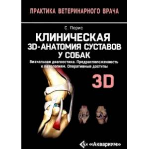
Clinical 3D anatomy of joints in dogs. Visual diagnosis. Distribution to pathologies
Please sign in so that we can notify you about a reply
Clinical 3D anatomy of joints in dogs. Visual diagnosis. Distribution to pathologies. Operational accesses
This book was written with the aim of solving the main problems that veterinary doctors face in the treatment of pathologies in the key joints of the limbs of dogs. Each joint is described in detail and presented in the smallest details: first, information on the anatomical organization of the joint is given, then detailed visual diagnostics, accompanied by three -dimensional drawings (radiography, simple or three -dimensional CT, CT in combination with color angiography, MRI)
The following is a detailed description of the most common pathologies of each joint, which are also accompanied by three -dimensional illustrations, radiographs, photographs of operations and other useful details.
Various approaches in the treatment of joint pathologies are also described in detail, also illustrated by photographs shot during anatomical preparation, which is very valuable because it gives a realistic idea of the procedures. Especially important are the surgical accesss presented in layers indicating the vascular-frozen trunks.
This publication will be useful for veterinarians, especially traumatologists and orthopedists. It can also be recommended as additional literature for students of veterinary universities in the disciplines: "Anatomy of animals", "clinical anatomy of animals", "Operational surgery with topographic anatomy of animals", "reconstructive surgery"
Author:
Author:Peris Salvador Clement
Cover:
Cover:Hard
Category:
- Category:Medical Books
Series:
Series: Practice of a veterinarian
ISBN:
ISBN:978-54238-0367-4
No reviews found
