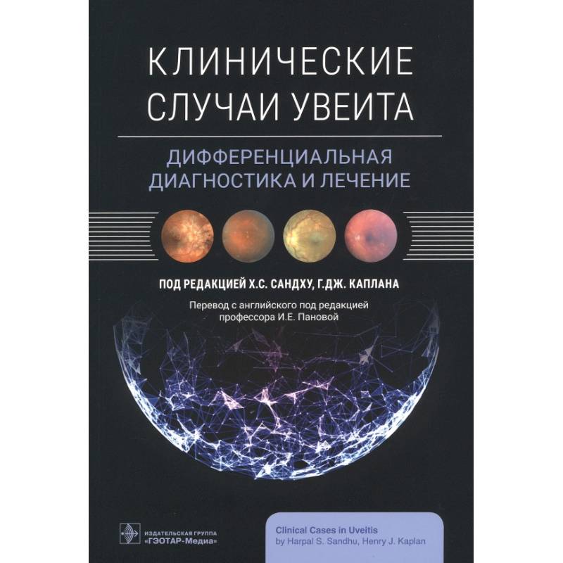
Clinical cases of uveitis. Differential diagnosis and treatment
Please sign in so that we can notify you about a reply
Intraocular inflammation is especially difficult to diagnose and treat, it often resembles a complex puzzle consisting of a patient"s medical history, symptoms, instrumental research and laboratory tests. This publication will help the doctor navigate the spectrum of complex manifestations and symptoms that often imitate various diseases. In the book in a simple format for understanding, more than 90 real cases of uveitis are presented, a step -by -step guide to collect an anamnesis, evaluation, differential diagnosis, testing, maintenance and subsequent observation is given.
Various cases of uveitis, scenarios for the development of diseases, as well as unique clinical situations found in everyday practice are given.
Contemporary methods of diagnostic visualization are described, including optical coherent tomography, optical coherent tomography-angiography, fluoresceine angiography and angiography with an Indian green. The features of diagnostic and methodological algorithms are presented, which help in differential diagnosis and decision making in the most difficult cases, including when the patient’s condition does not improve, as expected, which encourages the reassessment of the diagnosis and conduct of the disease.
The difference in infectious and non -infectious inflammation is discussed, when and how to start systemic immunosuppressive therapy, diagnostic criteria and treatment of white points syndrome, children"s uveit, masquerade syndromes and other issues. Contains more than 250 high-quality images, including color photographs of the front segment, fundus, octum imbalance and angiograms.
The publication is intended for practicing ophthalmologists and ophthalmic surgeons, general practitioners, clinical residents and graduate students of medical universities
Cover:
Cover:Soft
Category:
- Category:Science & Math
Publication language:
Publication Language:Russian
Paper:
Paper:Molded
ISBN:
ISBN:978-5-9704-7500-3
No reviews found
